Imaging-based Computational Biomedicine

Research Staff
-
Professor
Yoshinobu SATO -
Assistant Professor
Mazen SOUFI
| yoshi@is.naist.jp msoufi@is.naist.jp |
|
| To the site | http://icb-lab.naist.jp/index.html |
Research Areas
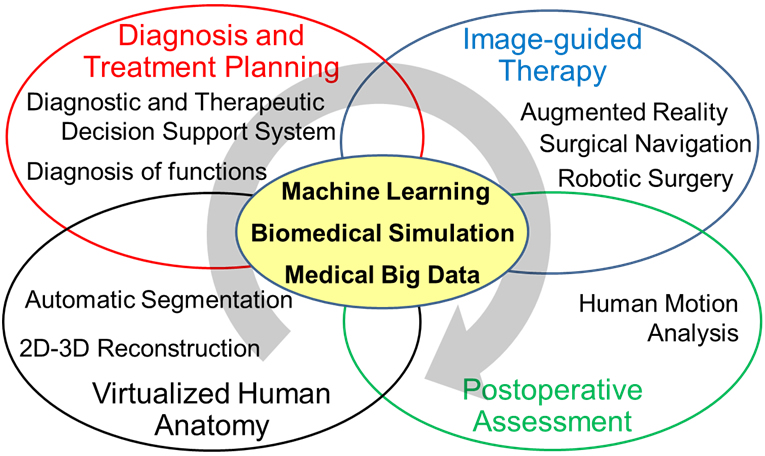
Fig. 1 Research areas in our lab
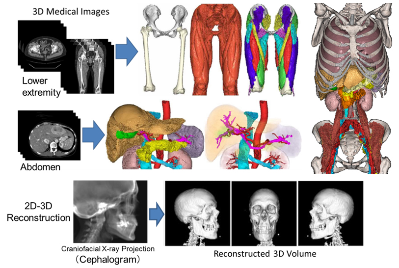
Fig. 2 Virtualized human anatomy
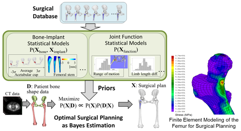
Fig. 3 Diagnosis and treatment planning
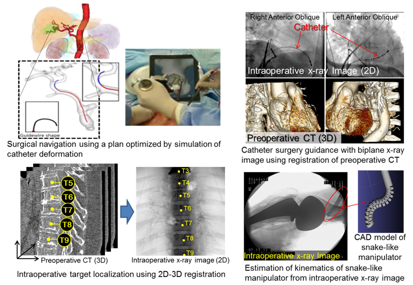
Fig. 4 Image-guided therapy
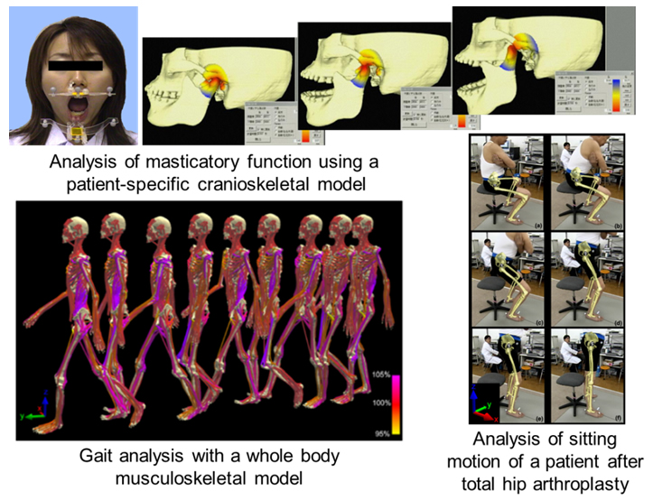
Fig. 5 Postoperative assessment
We integrate biomedical imaging with information science approaches such as machine learning including deep learning, computational simulation, and augmented reality to create knowledge and foster innovation in the field of computational biomedicine. We currently have four main research areas (Fig. 1):
Virtualized human anatomy (Fig. 2)
We create models of human anatomy for each subject from 3D biomedical images using AI technology. By integrating 3D image analysis and machine learning, we also create models of variability in anatomical shape and image appearance throughout a population, which we call computational anatomy models. We further construct computational models of, for example, physical or physiological functions to seek comprehensive understanding of a subject's body.
Diagnosis and treatment planning (Fig. 3)
We develop systems to support critical decision-making in diagnosis and therapeutic planning. These systems integrate patient-specific biomedical simulations with human anatomy models and statistical (or AI-based) predictions based on clinical databases (known as “medical big data”).
Image-guided therapy (Fig. 4)
We are developing a surgical navigation system to provide surgeons with intraoperative guidance throughby real-time fusion of the surgical field and surgical plans on patient anatomy models. Our goal is to develop "intelligence" in surgery based on statistical learning and computational simulations. This will enable the predictionng ofthe changing conditions of a patients during an operations in order to perform optimal surgical procedures.
Postoperative assessment (Fig. 5)
Medical treatment quality assurance requires proper assessment of the surgical outcomes. We develop methods to quantitatively evaluate the motion of patients who have had surgery on their musculoskeletal structures, such as in orthopedic and craniofacial operations, where detecting subtle changes in locomotion is crucial in predicting long-term outcome.
Key Features
Our laboratory features a highly integrated research environment for information science, biomedical imaging, clinical medicine, and other related technologies. We have a number of medical and technical collaborators, including companies, working together within Japan and throughout the world. We fully utilize our unique environment and our network of researchers to pursue our work in imaging-based computational biomedicine.